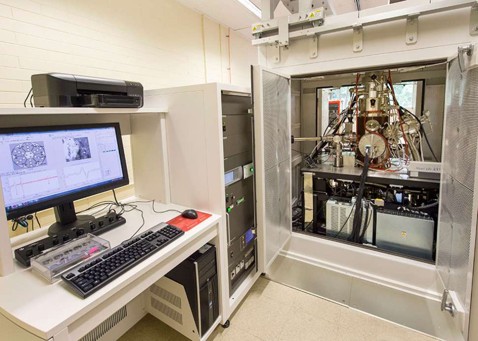
Scanning Auger Nanoprobe
The Scanning Auger Nanoprobe is one of only two in Australia and is able to map chemical information across a surface. This instrument combines high resolution electron microscopy with the ability to determine elemental composition, resulting the analysis of surface chemistry with a spatial resolution of 10 nanometres. Due to the nature of the species detected on the surface, the Scanning Auger Nanoprobe is very surface sensitive and able to detect low atomic number materials.
Contact
Instrument Leader: Professor Sarah Harmer
Scanning Auger Nanoprobe Facility Manager: Danielle Hughes
PHI 710 Scanning Auger Nanoprobe DOI: https://doi.org/10.25957/flinders.aes.phi710

- PHI 710 Scanning Auger Nanoprobe
- Field Emission Electron Source
- Secondary electron detector
- Cylindrical Mirror Electron Energy Analyser – reduces shadowing.
- Argon Ion sputter source -10kv-5000kv
Samples must be solid, dry and conductive. Sample dimensions up to 50mm x 50mm x 20mm
Application examples


Left: SEM image of Few-layer black phosphorous
Right: Overlayed Auger Electron maps of phosphorous (red), silicon (blue) and carbon (black), obtained at the same location.
From DOI: 10.1002/smtd.201700260


Left: SEM image of N2 plasma treated Graphene-Oxide. Right: Carbon Auger Electron elemental map obtained at the same location.
From DOI: 10.1039/C6CC04032B


Left: SEM image mild steel sample exposed to molten aluminium. Right: Overlayed Auger Electron elemental maps of iron (red) and aluminium (blue). Areas that show up purple indicate a mixture (eutectic) where aluminium has intruded into the steel surface.
From DOI: 10.1016/j.est.2018.03.001


Left: SEM image of a polished 3D printed 316 stainless steel surface. Right: Overlayed Auger Electron elemental maps of chromium (red), iron (green) and nickel (blue), taken at the same location.
Our equipment is funded by:


![]()
Sturt Rd, Bedford Park
South Australia 5042
South Australia | Northern Territory
Global | Online
CRICOS Provider: 00114A TEQSA Provider ID: PRV12097 TEQSA category: Australian University


